混合型拉曼系統
將 Renishaw 拉曼顯微鏡與各種製造商的其他分析系統結合,以提升其分析能力。
您可以用兩種以上的技術分析樣品,無須在兩種儀器之間奔波,進而達到最大效率。
利用 Renishaw 的關聯顯微系統,您可以放心地以兩種技術分析同一點。
聯絡我們
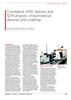
SPM/AFM:奈米解析度
將 inVia™ 拉曼顯微鏡結合原子力顯微鏡 (AFM) 等掃描測頭顯微鏡 (SPM),可用來研究材料的化學與結構特性。其中以針尖增強拉曼光譜 (TERS) 增添奈米級化學解析度,並揭露機械特性等補充資訊。
閱讀 Microscopy and Analysis Journal 中的文章
-
 Correlative SPM Raman and SEM analysis of biomedical devices and coatings [en]
A variety of medical implants and microelectrode arrays for electrophysiology are fabricated in thin film and micro technology. To guarantee the quality, proper functionality and safe operation of these devices, analytical techniques to investigate the structure and chemical composition of surfaces and interfaces during the fabrication process and for final quality control are essential.